Leg Bones Diagram : Feet Human Anatomy Bones Tendons Ligaments And More : At the same time, the bones and joints of the leg and foot must be strong enough to support the body's weight while remaining.
Leg Bones Diagram : Feet Human Anatomy Bones Tendons Ligaments And More : At the same time, the bones and joints of the leg and foot must be strong enough to support the body's weight while remaining.. Its lower end helps create the knee joint. The human leg, in the general word sense, is the entire lower limb of the human body, including the foot, thigh and even the hip or gluteal region. This page is about leg bones diagram,contains aluminium plant safety: Leg bones diagram / muscles that lift the arches of the feet | ankle anatomy. The hip joint is the uppermost part of the leg where the head of the thigh bone (femur) fits into the socket of the pelvis.
The hip joint is the uppermost part of the leg where the head of the thigh bone (femur) fits into the socket of the pelvis. Start studying cat leg bone anatomy. The horse leg anatomy in the rear includes the bones of the pelvis the ilium ischium and. The femur, or thigh bone, is the single bone of the thigh region (figure 6.51). Long bones are found in the arms (humerus, ulna, radius) and legs (femur, tibia, fibula), as well as in.
Free Vector Human Knee Anatomy Diagram from image.freepik.com Another bone that is part of the lower leg and the knee joint is called the fibula.this is a bone located on the lateral, or outer part, of the lower leg and is more commonly known as the calf bone. The pubis, ischium, and ilium together constitute the pelvis while the thigh bone is the femur. Separate the knee joint using a knife. It also separates muscles on the anterior and posterior parts of the leg. The following 29 files are in this category, out of 29 total. When you stand or walk, all the weight of your upper body rests on them. Most of the leg skeleton has bony prominences and margins that can be palpated and some serve as anatomical landmarks that define the extent of the leg. The thigh bone, or femur, is the large upper leg bone that connects the lower leg bones (knee joint) to the pelvic bone (hip joint).
The bones together make up the hip.
The major bones of the leg are the femur (thigh bone), tibia (shin bone), and adjacent fibula, and these are all long bones. Now let's look at the tibia bone, which is the larger of the two leg bones, located medially. Bone on side of the foot The lower leg extends from the knee to the ankle. Human knee anatomy diagram free vector. For the wings, hold the last 2 pinions so the exposed joint is uppermost and cut around the 11 lay out the chicken skin side down on a board, feel over the meat for any bones or cartilage and remove. The legs and feet the legs appear short when compared with the length of the body but they are powerful. Blank leg bones diagram : The bones of the leg are the femur tibia fibula and patellathe foot bones shown in this diagram are the talus navicular cuneiform cuboid metatarsals and calcaneus. The bones of the leg are the femur, tibia, fibula and patella.the foot bones shown in this diagram are the talus, navicular, cuneiform, cuboid, metatarsals and calcaneus. Upper leg bones diagram : The thigh bone, or femur, is the large upper leg bone that connects the lower leg bones (knee joint) to the pelvic bone (hip joint). The following 29 files are in this category, out of 29 total.
Related posts of diagram of leg bones bone of pelvis pics. This is the diagram of knee leg bone diagram that you search. The bones of the leg are the femur, tibia, fibula and patella.the foot bones shown in this diagram are the talus, navicular, cuneiform, cuboid, metatarsals and calcaneus. The tibia and fibula are two long bones that run parallel to each other, forming the scaffold of the leg and providing attachment points for many muscles. The foot bones shown in this diagram.
Dog Leg Anatomy In Human Speak Ortho Dog from orthodog.com Another bone that is part of the lower leg and the knee joint is called the fibula.this is a bone located on the lateral, or outer part, of the lower leg and is more commonly known as the calf bone. He leg's main function in the human is for locomotion and support of the rest of the body. Horses have a small bone just below their front knees called the third metacarpal, or shin bone, which supports their whole weight even when galloping. Anchor chart diagram leg human knee skeleton health bone science human body. The lower leg is comprised of two bones, the tibia and the smaller fibula. Bone diagram forehead (frontal bone) nose bones (nasals) cheek bone (zygoma) upper jaw (maxilla) lower jaw (mandible) breast bone (sternum) upper arm bone (humerus) lower arm bone (ulna) thigh bone (femur) collar bone (clavicle) toe bones (phalanges) ankle bones (tarsals) kneecap (patella) shin bone The second largest bone in physique is the tibia, additionally known as the shinbone. The human leg, in the general word sense, is the entire lower limb of the human body, including the foot, thigh and even the hip or gluteal region.
Blank leg bones diagram :
Horses have a small bone just below their front knees called the third metacarpal, or shin bone, which supports their whole weight even when galloping. The foot bones shown in this diagram. It also separates muscles on the anterior and posterior parts of the leg. Its lower end helps create the knee joint. The foot bones shown in this diagram are the talus, navicular, cuneiform, cuboid, metatarsals and calcaneus. The major bones of the leg are the femur (thigh bone), tibia (shin bone), and adjacent fibula, and these are all long bones. Bone diagram forehead (frontal bone) nose bones (nasals) cheek bone (zygoma) upper jaw (maxilla) lower jaw (mandible) breast bone (sternum) upper arm bone (humerus) lower arm bone (ulna) thigh bone (femur) collar bone (clavicle) toe bones (phalanges) ankle bones (tarsals) kneecap (patella) shin bone The femur, or thighbone, is the longest and largest bone in the human body. Leg bone diagram labeled : The foot bones shown in this diagram are the talus, navicular, cuneiform, cuboid, metatarsals and calcaneus. The rounded, proximal end is the head of the femur, which articulates with the acetabulum of the hip bone to form the hip joint. The bones together make up the hip. Separate the knee joint using a knife.
These muscles work together to produce movements such as standing, walking, running, and jumping. Long bones are found in the arms (humerus, ulna, radius) and legs (femur, tibia, fibula), as well as in. The bones of the leg are the femur, tibia, fibula and patella.the foot bones shown in this diagram are the talus, navicular, cuneiform, cuboid, metatarsals and calcaneus. It also separates muscles on the anterior and posterior parts of the leg. There also are bands of fibrous connective tissue—the ligaments and the tendons—in intimate relationship with the parts of the skeleton.
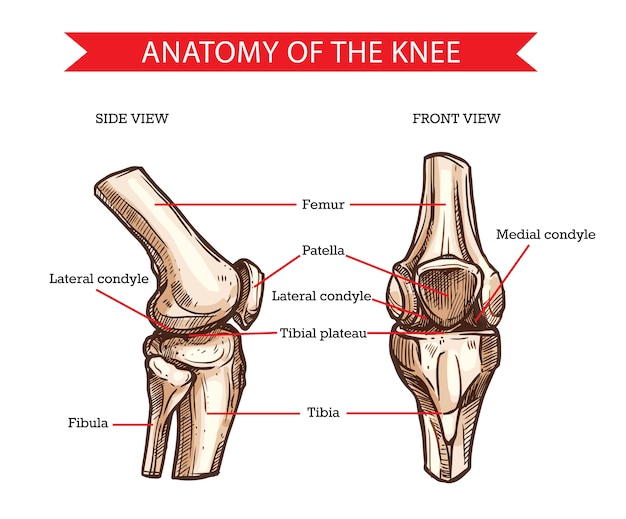
Premium Vector Anatomy Of Human Knee Sketch Of Leg Bones And Joint Medicine Side And Front View Of Knee Bones Hand Drawn Femur Patella Tibia And Fibula Tibial Plateau And from image.freepik.com For the wings, hold the last 2 pinions so the exposed joint is uppermost and cut around the 11 lay out the chicken skin side down on a board, feel over the meat for any bones or cartilage and remove. Its lower end helps create the knee joint. Bone of pelvis pics 12 photos of the bone of pelvis pics , bone. This is the diagram of knee leg bone diagram that you search. Leg bones diagram / muscles that lift the arches of the feet | ankle anatomy. The following 29 files are in this category, out of 29 total. Start studying cat leg bone anatomy. Bone on side of the foot
Includes leg (femur, tibia, patella, and fibula) and foot (tarsals and digits) bones.
Health diagram bone skeleton leg knee science anchor chart human human body. Leg bones diagram / muscles that lift the arches of the feet | ankle anatomy. The bones together make up the hip. The legs and feet the legs appear short when compared with the length of the body but they are powerful. The femur, or thighbone, is the longest and largest bone in the human body. The human leg, in the general word sense, is the entire lower limb of the human body, including the foot, thigh and even the hip or gluteal region. Includes leg (femur, tibia, patella, and fibula) and foot (tarsals and digits) bones. Upper leg bones diagram : Horses have a small bone just below their front knees called the third metacarpal, or shin bone, which supports their whole weight even when galloping. The lower leg extends from the knee to the ankle. Separate the knee joint using a knife. The foot bones shown in this diagram are the talus, navicular, cuneiform, cuboid, metatarsals and calcaneus. When you stand or walk, all the weight of your upper body rests on them.


0 Komentar